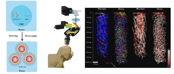
Scientists at Zhejiang University, including Prof. XI Wang and Prof. Anna Wang Roe at the Interdisciplinary Institute of Neuroscience and Technology and Prof. QIAN Jun at the College of Optical Science and Engineering, utilize a bright aggregation-induced emission (AIE) probe to achieve a large-depth three-photon fluorescence imaging of cortical microvasculature on nonhuman primates in vivo. Their research findings were published in the journal Biomaterials.
In humans and nonhuman primates, the structure of vascular organization is quite different from that in rodent. The cerebral cortex is functionally organized at mesoscale; that is, there are cortical columns of submillimeter sizes. Their clustered functional organization predicts that the oxygen and energetic demands are also necessarily clustered. Multiphoton microscopy can be suitable tools used for large-deep imaging at millimeter scales with literally no damage to the brain, thereby showing great promise for research into the dynamics of cerebral microvasculature. However, there are few studies in terms of three-photon fluorescence microscopy on the brain of nonhuman primates for lack of optimized imaging systems and excellent fluorescent probes.
Prof. XI Wang et al. firstly verified the bright AIE probe, marked with remarkable three-photon fluorescence efficiency and a convenient synthesis process, had no significant toxicity for nonhuman primates. They were then able to design and build a three-photon fluorescent microscopy system for the stable imaging of non-human primate brains. For the first time, three-dimensional maps of the cortical microvascular network were acquired with large depth up to 980 μm in a living macaque monkey. This might be the largest imaging depth obtained on the cortical microvasculature of macaque monkey by in vivo microscopic imaging methods at a micron resolution. Lastly, 3D reconstruction of the cortical microvascular network was performed, and similarities and differences between various parameters of brain structure between monkeys and mice were compared. This study shows that three-photon microscopy, as a primate-compatible method for imaging fine vascular networks, will facilitate the understanding of vascular functions in human brains.

The heart is often called the body’s “tireless pump,” working around the clock to keep us alive. But like any machine, it will wear down. When the heart becomes too weak to pump blood effectively o...
Cannabis has long walked a fine line between medicine and abuse. While it has shown potential for relieving pain and regulating mood, cannabis-based drugs are strictly controlled worldwide due to side...

A research article, entitled “More than microglial depletion: PLX5622 activates the hepatic constitutive androstane receptor to alter anesthesia and addiction” was published online in ...